8FHE | pdb_00008fhe

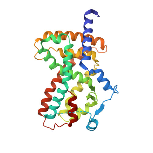
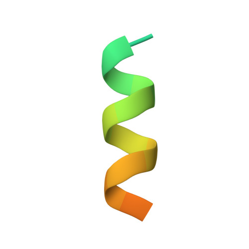
Crystal structure of PPARgamma ligand-binding domain in complex with N-CoR peptide and GW9662


Experimental Data Snapshot

  • Method: X-RAY DIFFRACTION
  • Resolution: 1.80 Å
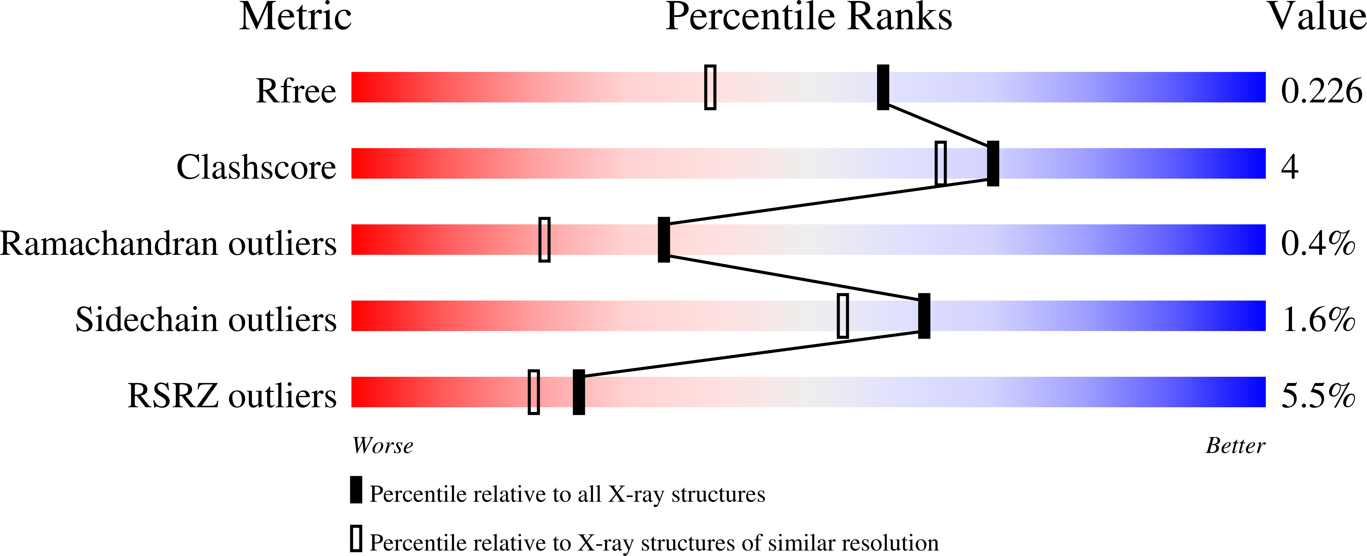
  • R-Value Free: 
    0.229 (Depositor), 0.230 (DCC) 
  • R-Value Work: 
    0.197 (Depositor), 0.200 (DCC) 
  • R-Value Observed: 
    0.199 (Depositor) 

Starting Model: experimental
View more details

wwPDB Validation   3D Report Full Report


Ligand Structure Quality Assessment 


This is version 1.2 of the entry. See complete history


Literature

Ligand efficacy shifts a nuclear receptor conformational ensemble between transcriptionally active and repressive states.

MacTavish, B.S.Zhu, D.Shang, J.Shao, Q.He, Y.Yang, Z.J.Kamenecka, T.M.Kojetin, D.J.

(2025) Nat Commun 16: 2065-2065

  • DOI: https://doi.org/10.1038/s41467-025-57325-4
  • Primary Citation of Related Structures:  
    8FHE, 8FHF, 8FHG, 8FKC, 8FKD, 8FKE, 8FKF, 8FKG

  • PubMed Abstract: 

    Nuclear receptors (NRs) are thought to dynamically alternate between transcriptionally active and repressive conformations, which are stabilized upon ligand binding. Most NR ligand series exhibit limited bias, primarily consisting of transcriptionally active agonists or neutral antagonists, but not repressive inverse agonists-a limitation that restricts understanding of the functional NR conformational ensemble. Here, we report a NR ligand series for peroxisome proliferator-activated receptor gamma (PPARγ) that spans a pharmacological spectrum from repression (inverse agonism) to activation (agonism) where subtle structural modifications switch compound activity. While crystal structures provide snapshots of the fully repressive state, NMR spectroscopy and conformation-activity relationship analysis reveals that compounds within the series shift the PPARγ conformational ensemble between transcriptionally active and repressive conformations that are natively populated in the apo/ligand-free ensemble. Our findings reveal a molecular framework for minimal chemical modifications that enhance PPARγ inverse agonism and elucidate their influence on the dynamic PPARγ conformational ensemble.


  • Organizational Affiliation

    Department of Integrative Structural and Computational Biology, Scripps Research and The Herbert Wertheim UF Scripps Institute for Biomedical Innovation & Technology, Jupiter, FL, USA.


Macromolecules
Find similar proteins by:  (by identity cutoff)  |  3D Structure
Entity ID: 1
MoleculeChains Sequence LengthOrganismDetailsImage
Peroxisome proliferator-activated receptor gammaA [auth B]275Homo sapiensMutation(s): 0 
Gene Names: PPARGNR1C3
UniProt & NIH Common Fund Data Resources
Find proteins for P37231 (Homo sapiens)
Explore P37231 
Go to UniProtKB:  P37231
PHAROS:  P37231
GTEx:  ENSG00000132170 
Entity Groups  
Sequence Clusters30% Identity50% Identity70% Identity90% Identity95% Identity100% Identity
UniProt GroupP37231
Sequence Annotations
Expand
  • Reference Sequence

Find similar proteins by:  Sequence   |   3D Structure  

Entity ID: 2
MoleculeChains Sequence LengthOrganismDetailsImage
Nuclear receptor corepressor 1B [auth D]23Homo sapiensMutation(s): 0 
Gene Names: NCOR1KIAA1047
UniProt & NIH Common Fund Data Resources
Find proteins for O75376 (Homo sapiens)
Explore O75376 
Go to UniProtKB:  O75376
PHAROS:  O75376
GTEx:  ENSG00000141027 
Entity Groups  
Sequence Clusters30% Identity50% Identity70% Identity90% Identity95% Identity100% Identity
UniProt GroupO75376
Sequence Annotations
Expand
  • Reference Sequence
Small Molecules
Ligands 1 Unique
IDChains Name / Formula / InChI Key2D Diagram3D Interactions
GW9 (Subject of Investigation/LOI)
Query on GW9

Download Ideal Coordinates CCD File 
C [auth B]2-chloro-5-nitro-N-phenylbenzamide
C13 H9 Cl N2 O3
DNTSIBUQMRRYIU-UHFFFAOYSA-N
Binding Affinity Annotations 
IDSourceBinding Affinity
GW9 BindingDB:  8FHE EC50: 2 (nM) from 1 assay(s)
Experimental Data & Validation

Experimental Data

  • Method: X-RAY DIFFRACTION
  • Resolution: 1.80 Å
  • R-Value Free:  0.229 (Depositor), 0.230 (DCC) 
  • R-Value Work:  0.197 (Depositor), 0.200 (DCC) 
  • R-Value Observed: 0.199 (Depositor) 
Space Group: P 41 21 2
Unit Cell:
Length ( Å )Angle ( ˚ )
a = 61.721α = 90
b = 61.721β = 90
c = 163.918γ = 90
Software Package:
Software NamePurpose
SCALAdata scaling
PHENIXrefinement
PDB_EXTRACTdata extraction
DIALSdata reduction
PHASERphasing

Structure Validation

View Full Validation Report


Ligand Structure Quality Assessment 


Entry History & Funding Information

Deposition Data


Funding OrganizationLocationGrant Number
National Institutes of Health/National Institute of Diabetes and Digestive and Kidney Disease (NIH/NIDDK)United States6R01DK124870

Revision History  (Full details and data files)

  • Version 1.0: 2024-03-20
    Type: Initial release
  • Version 1.1: 2024-10-23
    Changes: Structure summary
  • Version 1.2: 2025-04-30
    Changes: Database references