8HJ7 | pdb_00008hj7

Crystal structure of barley exohydrolase isoform ExoI E220A mutant in complex with beta-D-glucopyranose.


Experimental Data Snapshot

  • Method: X-RAY DIFFRACTION
  • Resolution: 1.85 Å
  • R-Value Free: 
    0.184 (Depositor), 0.193 (DCC) 
  • R-Value Work: 
    0.143 (Depositor), 0.157 (DCC) 
  • R-Value Observed: 
    0.145 (Depositor) 

Starting Model: experimental
View more details

wwPDB Validation   3D Report Full Report


Ligand Structure Quality Assessment 


This is version 1.1 of the entry. See complete history


Literature

The structure and dynamics of water molecule networks underlie catalytic efficiency in a glycoside exo-hydrolase.

Luang, S.Fernandez-Luengo, X.Streltsov, V.A.Marechal, J.D.Masgrau, L.Hrmova, M.

(2025) Commun Biol 8: 729-729

  • DOI: https://doi.org/10.1038/s42003-025-08113-9
  • Primary Citation of Related Structures:  
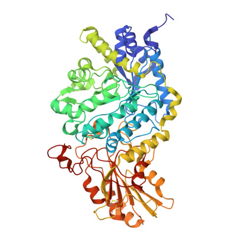
    8HJ6, 8HJ7, 8HJ8

  • PubMed Abstract: 

    Glycoside hydrolases break glycosidic bonds by transferring a water molecule onto the glycosidic oxygen of carbohydrates, but on the nanoscale, the dynamics of water molecules remains unclear. We investigate the role of the non-nucleophilic E220 glutamate, essential for maintaining the water molecule network in a family 3 β-D-glucan glucohydrolase, but not involved directly in catalysis. Kinetic data disclose that the E220A mutant retains substrate poly-specificity but has drastically reduced catalytic efficiency compared to the wild-type. High-resolution structures in-complex with a hydrolytic product and a mechanism-based inhibitor reveal that in wild-type, the concatenated water molecules near acid/base E491 and neighbouring N219 and E220 form a harmonised network. In contrast, in the E220A mutant, this network is uncoordinated. Computational models of covalent complexes show that water flux through the wild-type protein correlates with high catalytic efficiency dissimilar to E220A, where this correlation is lost. Ancestral sequence reconstructions of family 3 enzymes divulge the evolutionary conservation of residues participating in water molecule networks, which underlie substrate-product-assisted processivity. Our findings provide a blueprint for the dynamics of catalysis mediated by hydrolytic enzymes, which could inspire bioengineering to create a sustainable bio-economy.


  • Organizational Affiliation

    School of Agriculture, Food and Wine, and Waite Research Institute, Faculty of Sciences, Engineering and Technology, University of Adelaide, Adelaide, SA, Australia.


Macromolecules
Find similar proteins by:  (by identity cutoff)  |  3D Structure
Entity ID: 1
MoleculeChains Sequence LengthOrganismDetailsImage
Beta-D-glucan exohydrolase isoenzyme ExoI604Hordeum vulgareMutation(s): 1 
UniProt
Find proteins for Q9XEI3 (Hordeum vulgare subsp. vulgare)
Explore Q9XEI3 
Go to UniProtKB:  Q9XEI3
Entity Groups  
Sequence Clusters30% Identity50% Identity70% Identity90% Identity95% Identity100% Identity
UniProt GroupQ9XEI3
Glycosylation
Glycosylation Sites: 3
Sequence Annotations
Expand
  • Reference Sequence
Small Molecules
Ligands 6 Unique
IDChains Name / Formula / InChI Key2D Diagram3D Interactions
1PE (Subject of Investigation/LOI)
Query on 1PE

Download Ideal Coordinates CCD File 
O [auth A]PENTAETHYLENE GLYCOL
C10 H22 O6
JLFNLZLINWHATN-UHFFFAOYSA-N
NAG (Subject of Investigation/LOI)
Query on NAG

Download Ideal Coordinates CCD File 
B [auth A],
C [auth A],
D [auth A]
2-acetamido-2-deoxy-beta-D-glucopyranose
C8 H15 N O6
OVRNDRQMDRJTHS-FMDGEEDCSA-N
BGC (Subject of Investigation/LOI)
Query on BGC

Download Ideal Coordinates CCD File 
M [auth A],
N [auth A]
beta-D-glucopyranose
C6 H12 O6
WQZGKKKJIJFFOK-VFUOTHLCSA-N
SO4
Query on SO4

Download Ideal Coordinates CCD File 
P [auth A],
Q [auth A],
R [auth A],
S [auth A],
T [auth A]
SULFATE ION
O4 S
QAOWNCQODCNURD-UHFFFAOYSA-L
GOL (Subject of Investigation/LOI)
Query on GOL

Download Ideal Coordinates CCD File 
L [auth A]GLYCEROL
C3 H8 O3
PEDCQBHIVMGVHV-UHFFFAOYSA-N
ACT
Query on ACT

Download Ideal Coordinates CCD File 
E [auth A]
F [auth A]
G [auth A]
H [auth A]
I [auth A]
E [auth A],
F [auth A],
G [auth A],
H [auth A],
I [auth A],
J [auth A],
K [auth A]
ACETATE ION
C2 H3 O2
QTBSBXVTEAMEQO-UHFFFAOYSA-M
Experimental Data & Validation

Experimental Data

  • Method: X-RAY DIFFRACTION
  • Resolution: 1.85 Å
  • R-Value Free:  0.184 (Depositor), 0.193 (DCC) 
  • R-Value Work:  0.143 (Depositor), 0.157 (DCC) 
  • R-Value Observed: 0.145 (Depositor) 
Space Group: P 43 21 2
Unit Cell:
Length ( Å )Angle ( ˚ )
a = 100.634α = 90
b = 100.634β = 90
c = 182.321γ = 90
Software Package:
Software NamePurpose
REFMACrefinement
HKL-2000data reduction
HKL-2000data scaling
MOLREPphasing

Structure Validation

View Full Validation Report


Ligand Structure Quality Assessment 


Entry History & Funding Information

Deposition Data


Funding OrganizationLocationGrant Number
Other governmentAustralia--

Revision History  (Full details and data files)

  • Version 1.0: 2024-12-25
    Type: Initial release
  • Version 1.1: 2025-05-21
    Changes: Database references