9EMT | pdb_00009emt

Crystal structure of Histidine acetyltransferase with imidazole and coenzyme A disulfide


Experimental Data Snapshot

  • Method: X-RAY DIFFRACTION
  • Resolution: 1.40 Å
  • R-Value Free: 
    0.179 (Depositor), 0.180 (DCC) 
  • R-Value Work: 
    0.160 (Depositor), 0.159 (DCC) 
  • R-Value Observed: 
    0.161 (Depositor) 

Starting Model: experimental
View more details

wwPDB Validation   3D Report Full Report


Ligand Structure Quality Assessment 


This is version 1.2 of the entry. See complete history


Literature

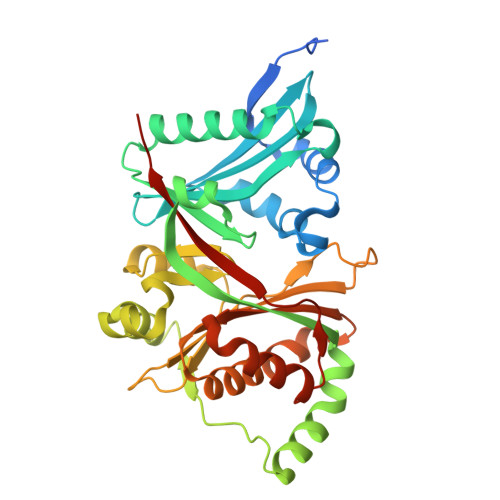
The molecular basis for acetylhistidine synthesis by HisAT/NAT16.

Myllykoski, M.Lundekvam, M.Osberg, C.Nilsen, S.S.Arnesen, T.

(2025) Nat Commun 16: 5960-5960

  • DOI: https://doi.org/10.1038/s41467-025-61145-x
  • Primary Citation of Related Structures:  
    9EMD, 9EMO, 9EMP, 9EMT, 9EN3

  • PubMed Abstract: 

    Acetylhistidine has been detected in human blood, but its origin and function are not known. It is formed when the acetyl group of acetyl-CoA is transferred to the α-amino group of histidine. Here we identify the intracellular NAT16 as the human histidine acetyltransferase (HisAT) responsible for histidine acetylation in vitro and in vivo. A NAT16 variant (p.Phe63Ser) present in over 5% of the population was previously found to correlate with reduced plasma levels of acetylhistidine and increased risk of kidney disease. Our biochemical analysis of HisAT/NAT16 Phe63Ser shows reduced affinity for Histidine supporting a model where this variant has less acetylhistidine catalysis leading to lower blood level of acetylhistidine. We find that HisAT adopts a double-GNAT (Gcn5-related N-Acetyltransferase) fold where the N-terminal domain binds acetyl-CoA and with distinct active site conformation allowing the binding of histidine in between the two domains. We detect similar structures from across living organisms and find that the HisAT structure is conserved in several archaeal and bacterial species. In sum, NAT16 is the human histidine acetyltransferase utilizing a rare double-GNAT structure to steer plasma acetylhistidine levels with potential impact for kidney function.


  • Organizational Affiliation

    Department of Biomedicine, University of Bergen, Bergen, Norway. [email protected].


Macromolecules
Find similar proteins by:  (by identity cutoff)  |  3D Structure
Entity ID: 1
MoleculeChains Sequence LengthOrganismDetailsImage
Probable N-acetyltransferase 16352Homo sapiensMutation(s): 0 
Gene Names: NAT16C7orf52
EC: 2.3.1
UniProt & NIH Common Fund Data Resources
Find proteins for Q8N8M0 (Homo sapiens)
Explore Q8N8M0 
Go to UniProtKB:  Q8N8M0
PHAROS:  Q8N8M0
GTEx:  ENSG00000167011 
Entity Groups  
Sequence Clusters30% Identity50% Identity70% Identity90% Identity95% Identity100% Identity
UniProt GroupQ8N8M0
Sequence Annotations
Expand
  • Reference Sequence
Small Molecules
Ligands 4 Unique
IDChains Name / Formula / InChI Key2D Diagram3D Interactions
5NG (Subject of Investigation/LOI)
Query on 5NG

Download Ideal Coordinates CCD File 
B [auth A][[(2~{S},3~{S},4~{R},5~{R})-5-(6-aminopurin-9-yl)-4-oxidanyl-3-phosphonooxy-oxolan-2-yl]methoxy-oxidanyl-phosphoryl] [(3~{R})-4-[[3-[2-[2-[3-[[(2~{R})-4-[[[(2~{R},3~{S},4~{R},5~{R})-5-(6-aminopurin-9-yl)-4-oxidanyl-3-phosphonooxy-oxolan-2-yl]methoxy-oxidanyl-phosphoryl]oxy-oxidanyl-phosphoryl]oxy-3,3-dimethyl-2-oxidanyl-butanoyl]amino]propanoylamino]ethyldisulfanyl]ethylamino]-3-oxidanylidene-propyl]amino]-2,2-dimethyl-3-oxidanyl-4-oxidanylidene-butyl] hydrogen phosphate
C42 H70 N14 O32 P6 S2
YAISMNQCMHVVLO-BJLFRJKCSA-N
MLI
Query on MLI

Download Ideal Coordinates CCD File 
E [auth A]MALONATE ION
C3 H2 O4
OFOBLEOULBTSOW-UHFFFAOYSA-L
GOL
Query on GOL

Download Ideal Coordinates CCD File 
D [auth A],
F [auth A],
G [auth A]
GLYCEROL
C3 H8 O3
PEDCQBHIVMGVHV-UHFFFAOYSA-N
IMD (Subject of Investigation/LOI)
Query on IMD

Download Ideal Coordinates CCD File 
C [auth A]IMIDAZOLE
C3 H5 N2
RAXXELZNTBOGNW-UHFFFAOYSA-O
Experimental Data & Validation

Experimental Data

  • Method: X-RAY DIFFRACTION
  • Resolution: 1.40 Å
  • R-Value Free:  0.179 (Depositor), 0.180 (DCC) 
  • R-Value Work:  0.160 (Depositor), 0.159 (DCC) 
  • R-Value Observed: 0.161 (Depositor) 
Space Group: P 63
Unit Cell:
Length ( Å )Angle ( ˚ )
a = 91.129α = 90
b = 91.129β = 90
c = 98.487γ = 120
Software Package:
Software NamePurpose
PHENIXrefinement
XDSdata reduction
XDSdata scaling
PHASERphasing
Cootmodel building

Structure Validation

View Full Validation Report


Ligand Structure Quality Assessment 


Entry History & Funding Information

Deposition Data


Funding OrganizationLocationGrant Number
European Research Council (ERC)European Union772039

Revision History  (Full details and data files)

  • Version 1.0: 2025-03-26
    Type: Initial release
  • Version 1.1: 2025-07-09
    Changes: Database references
  • Version 1.2: 2025-07-16
    Changes: Database references