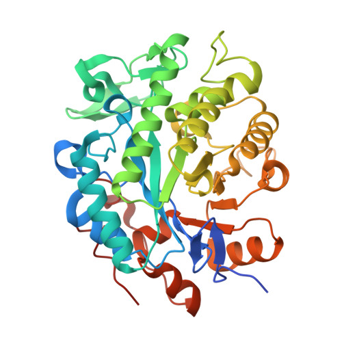
Rational Introduction of Electrostatic Interactions at Crystal Contacts to Enhance Protein Crystallization of an Ene Reductase.
Walla, B., Maslakova, A., Bischoff, D., Janowski, R., Niessing, D., Weuster-Botz, D.(2025) Biomolecules 15
- PubMed: 40305164
- DOI: https://doi.org/10.3390/biom15040467
- Primary Citation of Related Structures:
9QGB, 9QGC, 9QGD, 9QGE, 9QGF - PubMed Abstract:
Protein crystallization is an alternative to well-established but cost-intensive and time-consuming chromatography in biotechnological processes, with protein crystallization defined as an essential unit operation for isolating proteins, e.g., active pharmaceutical ingredients. Crystalline therapeutic proteins attract interest in formulation and delivery processes of biopharmaceuticals due to the high purity, concentration, and stability of the crystalline state. Although improving protein crystallization is mainly achieved by high-throughput screening of crystallization conditions, recent studies have established a rational protein engineering approach to enhance crystallization for two homologous alcohol dehydrogenases from Lactobacillus brevis ( Lb ADH) and Lactobacillus kefiri ( Lk ADH). As generalizing crystallization processes across a wide range of target proteins remains challenging, this study takes a further step by applying the successful crystal contact engineering strategies for Lb ADH/ Lk ADH to a non-homologous protein, an NADH-binding derivative of the Nostoc sp. PCC 1720 ene reductase ( Nsp ER1-L1,5). Here, the focus lies on introducing electrostatic interactions at crystal contacts, specifically between lysine and glutamic acid. Out of the nine tested Nsp ER1-L1,5 mutants produced in E. coli , six crystallized, while four mutants revealed an increased propensity to crystallize in static µL-batch crystallization compared to the wild type: Q204K, Q350K, D352K, and T354K. The best-performing mutant Q204K was selected for upscaling, crystallizing faster than the wild type in a stirred batch crystallizer. Even when spiked with E. coli cell lysate, the mutant maintained increased crystallizability compared to the wild type. The results of this study highlight the potential of crystal contact engineering as a reliable tool for improving protein crystallization as an alternative to chromatography, paving the way for more efficient biotechnological downstream processing.
- Biochemical Engineering, Department of Energy and Process Engineering, TUM School of Engineering and Design, Technical University of Munich, Boltzmannstraße 15, 85748 Garching, Germany.
Organizational Affiliation:


